Pseudoaneurysm of the Mitral-aortic Intervalvular Fibrosa: An Update
In February 2010, I issued a call for cases of pseudoaneurysm of the mitral-aortic intervalvular fibrosa (MAIVF). Now, one year later, I think it is appropriate to suspend the project. First of all, the condition is so rare that only one other case was submitted to add to my own collection. Secondly, a comprehensive review article was published in the Journal of the American Society of Echocardiography in October 2010. That article reviewed 88 patients from the English-language literature published from 1966 to the end of 2009. The author added his own case for a total of 89 cases. Such a review renders our efforts unnecessary.
Here is what they report: pseudoaneurysm of the mitral-aortic intervalvular fibrosa (P-MAIVF) is a rare condition that has been reported to occur following endocarditis and surgical trauma. This is an entity that occurs in the interannular zone between the mitral and aortic valves and its communication with the left ventricular outflow tract between the left coronary or non-coronary aortic cusp and the anterior leaflet of the mitral valve.
Three-quarters of patients present with symptoms and signs of infection, chest pain, heart failure or shortness of breath and cerebrovascular accidents. The age range is between 3 months and 90 years. About half of the patients had prosthetic aortic valves and the clear majority of those valves were mechanical as opposed to bioprosthetic. Of the patients with this complication and native valves, 33% of those were bicuspid valves.
Active or prior endocarditis was present in 64 patients and there was no endocarditis in 19. Surgery is considered to be the treatment of choice. It was performed in 68 patients. Fifty-six had aortic valve replacement with P-MAIVF repair. Ten patients had P-MAIVF repair with no valve surgery and one had P-MAIVF and mitral valve repair.
Of the 8 patients who did not undergo surgery, one went to hospice for palliative care. In one patient with four year follow up, the pseudoaneurysm increased in size and in the other two there were no changes at 10 month and 3 year follow up. (Of my patients, the first had spontaneous thrombosis and resolution of the P-MAIVF and the other went to repeat mitral valve surgery because of infective endocarditis.) Fourteen patients died: 3 from P-MAIVF rupture into the pericardium, 3 from sepsis, and 6 post operatively. One had an unrelated drug overdose. One patient with no medical history had sudden death and autopsy showed changes consistent with compression of the left circumflex artery. (We saw a case of compression of the circumflex by P-MAIVF at the Echo in the Slopes meeting this past January.)
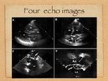
Theodore M. Batzer, M.D. of Manistee, Michigan submitted a great case for our series. A still frame of that study is posted here as figure 1.
Felix J. Rogers, D.O.
FIGURE 1: Pseudoaneurysm of the mitral aortic intervalvular fibrosa. The patient had replacement of the aortic valve with a bioprosthetic device in December 2001 with mitral valve repair. He has had several significant infections including septic risk (staph aureus about 2005) and the infected total knee arthroplasty with enterococcus faecalis (not VRE) in 2006 with a subsequent re-do total knee arthroplasty that year.
|